- Organic Geochemistry
- Laboratories
- GBM Imaging Laboratory
Geobiomolecular Imaging Laboratory
Introduction
In the framework of the ZOOMecular project, the Geobiomolecular Imaging Lab was established at the University of Bremen to implement mass spectrometry imaging techniques (MSI) in the geosciences. Conventional analysis of molecular biomarkers and proxies is limited in spatial and temporal resolution due to the need of ~cm3 sized bulk samples. Our innovative MSI approach enables their analysis with ultra-high (sub-mm) resolution directly on the surface of geological samples. Our expertise includes the preparation of samples, the spatially resolved analysis of molecular biomarkers and complementary data, and the evaluation and visualization of obtained maps of biomarker distributions. A major field of application for this technique are paleoclimate and paleoenvironmental reconstructions based on molecular proxies archived in the sedimentary record. With the sub-mm resolution offered by MSI, such reconstructions can be obtained with unprecedented resolution, and even seasonal variability can be accessed.
The analytical centerpiece of the Geobiomolecular Imaging Lab is a 7T solariX XR FT-ICR-MS equipped with a MALDI source that provides excellent mass accuracy, resolveing power and sensitivity and is therefore ideally suited for mapping molecular biomarkers and proxies in geological samples. Additionally, elemental distributions in the same sample can be mapped via Micro-XRF spectrometry (µ-XRF).
Laser-desorption ionization (LDI) coupled to ultra-high resolution mass spectrometry (FTMS)
MALDI-based MSI relies on the laser-induced desorption of the analyte from the sample, its subsequent primary ionization by the laser pulse and secondary ionization in the expanding ion cloud, and detection by mass spectrometry. With the laser rastering across the surface of the sample in a defined submillimeter-fine grid, target molecules can be locally resolved. The coupling to Fourier transform ion cyclotron resonance mass spectrometry (FT-ICR-MS) provides high sensitivity and accurate identification of informative compounds.

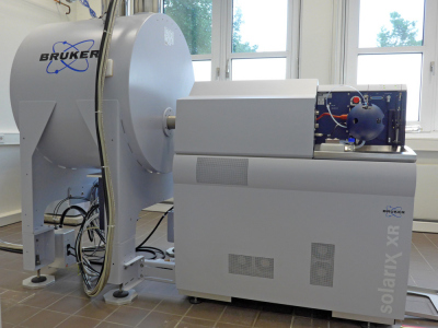
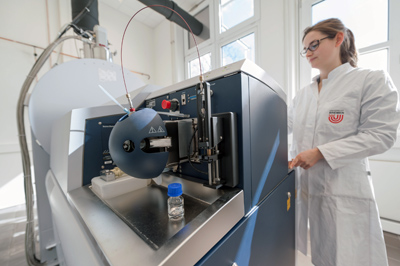
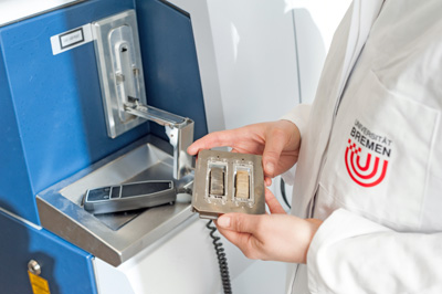
Bruker solariX FTMS with LDI
Our FTMS system is a 7T solariX XR FT-ICR-MS coupled to a MALDI source equipped with a Smartbeam II laser (Bruker Daltonik, Bremen).
Specifications:
Superconducting 7T magnet
Basic assembly
Sample introduction devices
Vacuum system for mass spectrometer
Provision for ion transfer to the detector and ion manipulation
to the analyzer (ICR) cell
NanoBay Console
MALDI user access area
FT-ICR mass spectrometer

Micro-XRF spectrometry (µ-XRF)
Principles of Micro-XRay Fluorescence-Spectrometry
µ-XRF is based on the excitation of material with X-ray radiation and detection of the emitted fluorescence radiation spectrum whereby each element reacts at characteristic energy lines. The fluorescence yield is growing with atomic number and can be enhanced by generating vacuum in the instrument chamber if dry samples are measured. Elements are detected by fluorescence emittance in total of all their chemical forms.
µ-XRF scanning works as non-contact and non-destructive measurement on sample surfaces. For maximal detection yields, sample surfaces are plain, not necessarily polished. Depending on the kind of material (metal – mineral – organic) and on the atomic number, the sample is investigated through the surface to a depth ranging from µm to mm. Scanning methods of the M4 Tornado µ-XRF are point measurement, line scanning and area mapping. During an area mapping, a full element spectrum is detected for each pixel in the map. Measurements are possible in high resolution down to 5 µm pixel size using polycapillary optics.
Research interests
Elemental mapping on sediment surfaces (at high spatial resolution) to characterize the biogeochemical microenvironment in studying biomarker-mineral association
Characterization of geological samples (bulk and thin sections)
Investigating microbial mats, chimneys and other objects

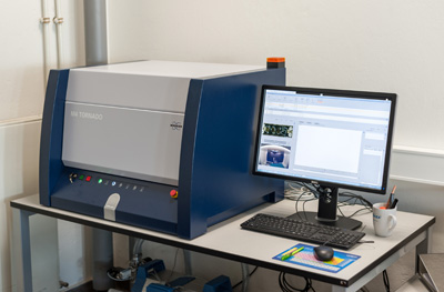
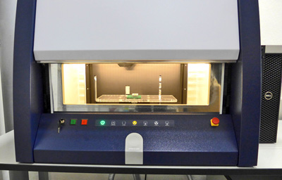
Bruker M4 Tornado 300 µ-XRF
The benchtop µ-XRF scanner is equipped with two optics, a polycapillary and a collimator.
Specifications:
HV-generator max. 50kV
X-ray tube 1: Microfocus Rh-anode and Poly capillary optic (20 µm spot)
X-ray tube 2: Feinfocus W-anode and Collimator optic (1 mm spot)
XFlash® Silicon Drift Detector 30 mm²
Turbospeed X-Y-Z-sample stage
Primary filter set
Vacuum pump set
Video cameras 10x, 100x

Sample preparation
The cryomicrotome is used to cut the embedded frozen samples into slices of sub-mm thickness.
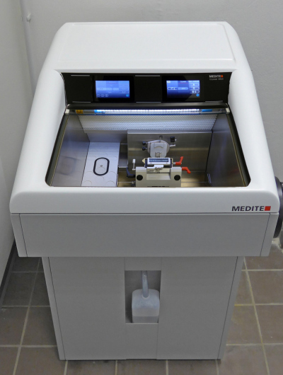
Medite Cryostat M630
The semi-automatic Cryostat M630 can cool down the temperature range to -35°C of the microtome and cutting work area.
Specifications:
Flash freezing unit
Fast freezing stage for specimen
Sectioning range: 1 µm to 100 µm in 1 µm steps
Trimming range: 5 to 100 µm in 5 µm steps and 100 to 500 µm in 50 µm steps
High precision motorized coarse feed movement
Automatic defrosting function

The Bruker solariX LDI FTMS was funded by
“The European Research Council (ERC) under the European Union’s Horizon 2020 research and innovation programme (grant agreement No 670115)”
