Page path:
- Modern Dinocyst Key
- brown cysts
- spherical brown randomly distributed processes
- spherical brown cysts with hollow processes
- Cyst of Protoperidinium fukuyoi
Cyst of Protoperidinium fukuyoi
Zonneveld, K.A.F. and Pospelova V. (2015). A determination key for modern dinoflagellate cysts. Palynology 39 (3), 387- 409.
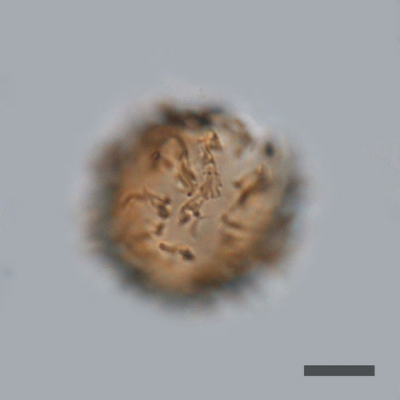
lateral view
Photographs by Vera Pospelova
locality: Saanich Inlet (Canada)
Photographs by Vera Pospelova
locality: Saanich Inlet (Canada)

cross section
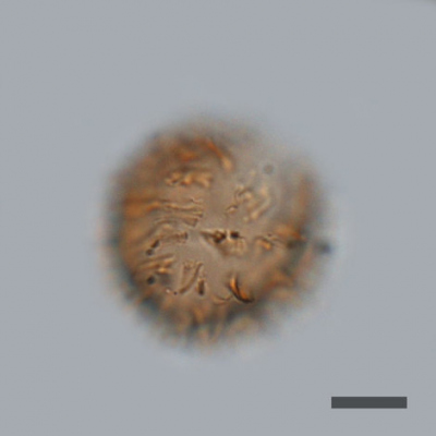
lateral view
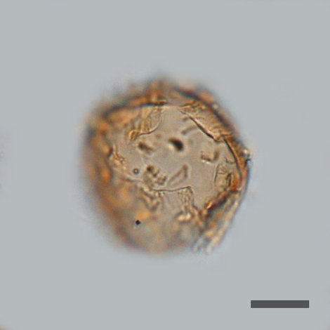
dorsal view
Photographs by Vera Pospelova
locality: Saanich Inlet (Canada)
Photographs by Vera Pospelova
locality: Saanich Inlet (Canada)
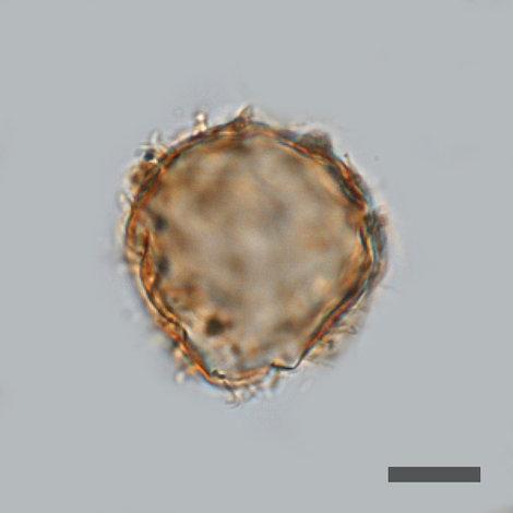
cross section
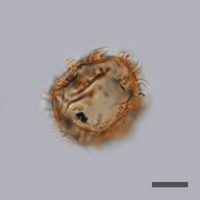
dorsal view
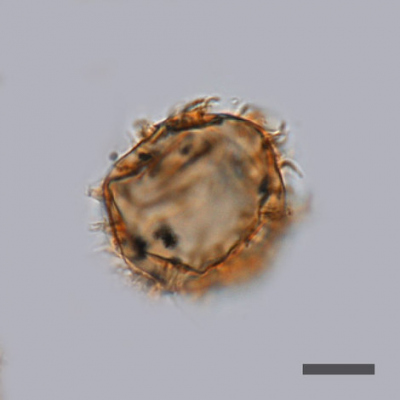
cross section
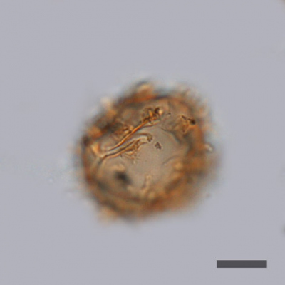
ventral view
Photographs represent figures 22, 24 and 30 from: Mertens et al., (2013) Journal of eukaryotic Microbiology, 0, 1-19. doi: 10.1111/jeu.12058
Field characteristics
Cyst of Protoperidinium fukuyoi Mertens et al., 2013
Characteristics:
Small light brown to golden brown cysts with smooth to finely granular cyst wall covered by numerous processes. Processes are hollow with circular bases that distally become flattened and bladelike. Processes typically grouped into clusters with adjacent processes in each cluster fusing at their bases and often for much or most of their length. Distal ends of processes are typically free. Clusters are arranged in straight or curved lineations. Process length is fairly constant for individual specimens, but varies between specimens. Occasional scattered hairlike processe processes up to 0.3 µm can be present. Apical is formed by the loss of the 2th, 3th and 4th apical plates as well as the pore plates.
Dimensions: Cyst body diameter: 44 to 48 µm; length of processes: 5 to 8 µm.
Motile affinity: unknown
Stratigraphic range: Pleistocene to recent
Comparison with other species:
This species differs from all other species in having processes that become distally flattened and ribbon-like. Furthermore, processes are grouped in clusters.


