Page path:
- Modern Dinocyst Key
- brown cysts
- brown elongate cysts
- Cyst of Gymnodinium trapeziforme
Cyst of Gymnodinium trapeziforme
Zonneveld, K.A.F. and Pospelova V. (2015). A determination key for modern dinoflagellate cysts. Palynology 39 (3), 387- 407.
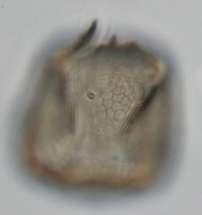
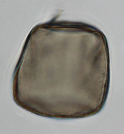
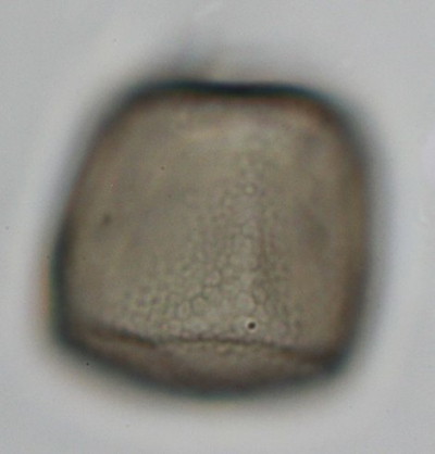
lateral view
sample GeoB 12312-2
region: Arabian Sea Makran OMZ
phtographs: Karin Zonneveld
sample GeoB 12312-2
region: Arabian Sea Makran OMZ
phtographs: Karin Zonneveld
cross section
lateral view
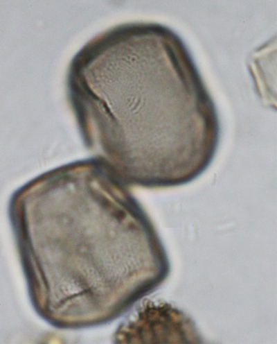
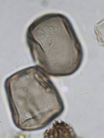
lateral view
sample GeoB 12312-2
region: Arabian Sea Makran OMZ
phtographs: Karin Zonneveld
sample GeoB 12312-2
region: Arabian Sea Makran OMZ
phtographs: Karin Zonneveld
lateral view
Field characteristics
Cyst of Gymnodinium trapeziforme Attaran Fariman and Bolch 2007
Field characteristics:
Trapezoidal to subrectangular pale-brown to purple-brown cysts that are dorsoventrally compressed. Epicyst is often somewhat smaller than hypocyst. Cysts are covered with a polygonal reticulum that reflect cingulum, sulcus and amphiesmal vesicles of the vegetative cell. Cingulum width is about one fourth of the cyst length. Cingulum is reflected by two double rows of vesicles with three to four rows of paravesicles between the margins. The sulcus is reflected by a double row of paravesicles arranged in a straight line extending from near the antapex up to the ventral pore region. A single row of rectangular paravesicles extend from the flagellar pore to the apex. The apical groove is not clearly reflected. In the flagellar pore area the paravesicles are larger in size and irregularly shaped. Archeopyle chasmic with varying orientation.
Dimensions: Length 23 to 34 µm (26 µm), width 17 to 28 µm (21 µm) with 2 to 7 µm difference between the narrowest and widest point.
Cyst theca relationship: Attaran Fariman and Bolch, 2007
Stratigraphy: Recent
Comparison with other species:
This cyst is easy to distinguish as it is the only presently described cyst that has a micro reticulate brown cyst that is not spherical in outline.
Personally (Karin) I have seen high concentrations of these cysts in sediments from the Pakistan shelf oxygen minimum zone where they can dominate the assemblage.


